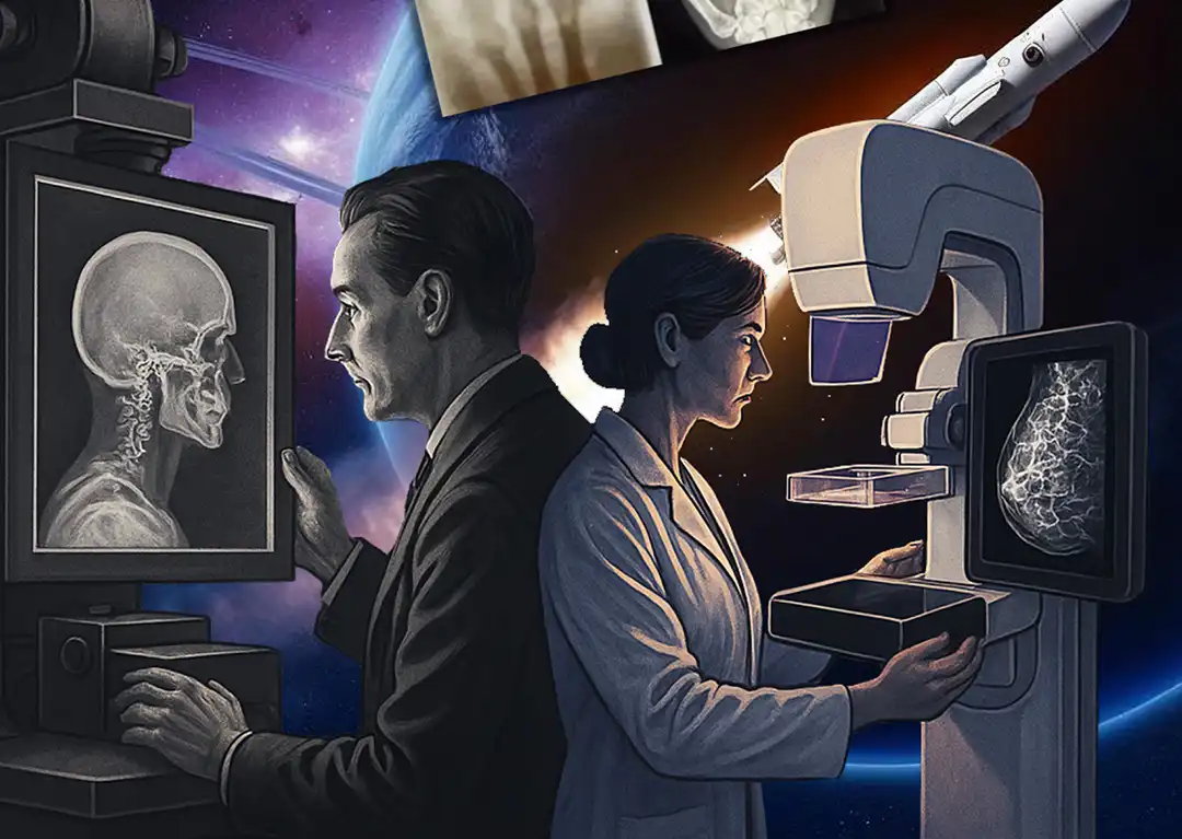
The Evolution of X-ray in Mammography and Beyond
In 1895, Wilhelm Röntgen’s accidental discovery of X-rays marked a transformative moment in medical history. While experimenting with vacuum tubes, he noticed that mysterious “new rays” could pass through solid objects and cast shadows—famously capturing an image of his wife’s hand. This breakthrough quickly became a powerful diagnostic tool, reshaping the practice of medicine.
Early Milestones in Breast Imaging
The application of X-rays to breast imaging began in the early 20th century. In 1913, German surgeon Albert Salomonconducted the first mammographic studies using tissue removed during mastectomies. His pioneering work demonstrated how X-rays could distinguish between benign and malignant tumors and trace cancers spread through lymph nodes. While his research laid the foundation for modern mammography, it was limited to excised tissue.
In the 1930s, American radiologist Stafford Warren advanced the field by performing mammograms on living patients. He emphasized imaging both breasts for comparative analysis, a practice still followed today.
A pivotal innovation came in 1949 from Uruguayan radiologist Raul Leborgne, who introduced breast compression. This technique reduced radiation exposure while dramatically improving image quality, enabling the detection of subtle abnormalities such as microcalcifications—now recognized as key indicators of breast cancer.
Standardization and Technological Growth (1960s–1990s)
The 1960s and 1970s marked a period of standardization. Robert Egan developed consistent mammographic techniques using high milliamperage and low kilovoltage settings with specialized film. His methods improved diagnostic accuracy and facilitated wider adoption of mammography for breast cancer screening. Egan also showed that mammography could detect cancers not yet palpable during a physical exam.
Charles Gros designed the first dedicated mammography X-ray system in France in the late 1960s. His unit featured molybdenum tubes for enhanced soft tissue contrast and incorporated a built-in compression system.
Around the same time, Philip Strax conducted landmark clinical trials that proved the effectiveness of mammography in reducing breast cancer mortality, firmly establishing it as a key screening tool.
In the 1980s, innovations in screen-film technology improved image clarity while reducing radiation exposure. Xeromammography, which used a dry process for imaging, provided even greater detail.
The American College of Radiology (ACR) led efforts to standardize mammography practices and radiologist training. In 1993, the introduction of BI-RADS (Breast Imaging Reporting and Data System) provided a unified language for interpreting and reporting mammographic findings.
The Digital Revolution and Advanced Imaging (2000–Today)
The new millennium ushered in the era of digital imaging. In 2000, the FDA approved the first full-field digital mammography (FFDM) system, enabling instant image review and manipulation.
In 2011, digital breast tomosynthesis (DBT)—or 3D mammography—received FDA approval. DBT captures multiple images from different angles, reconstructing a 3D view of the breast. This reduces tissue overlap, especially in dense breasts, and improves detection rates while lowering false positives.
Recent advances include:
- Contrast-enhanced mammography (CEM) highlights areas of increased blood flow associated with tumors.
- Emerging technologies like molecular breast imaging (MBI) and breast-specific gamma imaging (BSGI) offer better visualization of dense breast tissue.
- The integration of artificial intelligence (AI) and radiomics promises more precise and personalized imaging diagnostics.
Pioneers Who Shaped the Field
The evolution of mammographic imaging is the result of contributions from many visionaries:
- Wilhelm Röntgen (1895): Discovered X-rays.
- Albert Salomon (1913): Conducted the first mammograms on mastectomy specimens.
- Stafford Warren (1930s): Pioneered mammography on live patients.
- Raul Leborgne (1949): Introduced breast compression.
- Helen Ingleby: Enhanced understanding of breast anatomy through histology.
- Robert Egan (1960s): Standardized imaging techniques.
- Charles Gros (1960s): Designed the first dedicated mammography system.
- Philip Strax (1966): Validated mammography’s value in large-scale screening.
- Other notable contributors include Price, Butler, Ostrum, Becker, Isard, Moskowitz, Sickles, Kopans, Homer, and Tabár, along with companies like Kodak and Dupont, which advanced imaging films and intensifying screens.
Then, there is Elizabeth Fleischmann. She is one of the earliest women in radiology, establishing her own X-ray lab in the late 19th century, helping to pave the way for diagnostic imaging despite personal risk and limited recognition.
A Leap Into Space: The First Medical X-Ray in Orbit
On April 2, 2025, aboard a SpaceX Dragon capsule, Fram2 Mission Commander Chun Wang captured the first-ever medical X-ray in space—a powerful symbol of medical progress. This achievement used specialized, portable imaging systems developed by KA Imaging and MinXray, designed for operation in microgravity.
The image—a hand with a ring—paid homage to Röntgen’s original X-ray. But this mission represented far more than symbolism. It validated that safe, efficient diagnostic imaging is possible in space, a critical capability for long-duration missions.
The technology, originally built for battlefield use, was adapted for space, highlighting how the extreme requirements of microgravity—miniaturization, safety, low power, and portability—can drive innovation with far-reaching applications. The KA Imaging detector’s ability to differentiate between bone and soft tissue in a single exposure is promising for astronauts and patients in remote and underserved communities on Earth.
Beyond medical diagnostics, the mission also showed how X-rays can support non-invasive equipment testing, enabling real-time troubleshooting on spacecraft.
Toward Interplanetary Healthcare
This “cosmic first” brings us closer to a future where comprehensive medical care—including screening mammograms, diagnostic ultrasounds, and advanced imaging—can be delivered even in deep space. As humans journey further from Earth, technologies like space-based X-rays will be essential to monitoring health, managing injuries, and ensuring mission success.
Just as Röntgen’s invisible rays once unlocked new frontiers in medicine, Commander Wang’s orbital X-ray marks a bold new chapter in human exploration—one where the pursuit of health, safety, and discovery extends beyond our planet.

