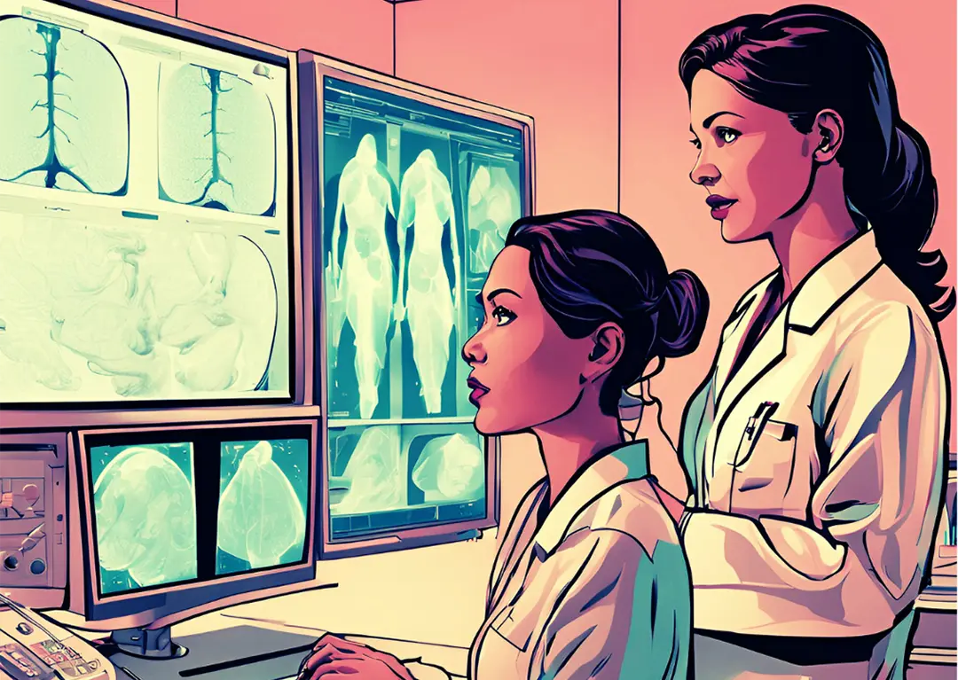
Breast cancer risk assessment is undergoing a revolution, driven by the convergence of traditional medical models and cutting-edge artificial intelligence (AI).
What if we could predict a woman’s breast cancer risk with unprecedented accuracy, not just based on her family history or age, but by analyzing patterns hidden deep within her mammograms?
This evolving landscape of risk prediction, where clinical expertise meets machine learning, promises to transform how we detect, prevent, and manage breast cancer and is no longer somewhere out there in the future. It is here now.
From the trusted Gail Model to AI-powered tools like Mirai and ProFound AI Risk, this article explores some of the questions about how these approaches compare and what lies ahead for personalized care.
Traditional Risk Assessment Models
The Gail Model
The Breast Cancer Risk Assessment Tool (BCRAT), commonly known as the Gail Model, was developed in 1989 by Dr. Mitchell Gail. It has since become one of clinical practice’s most widely used risk assessment tools. The Gail Model estimates a woman’s risk of developing invasive breast cancer within five years and up to age 90 based on factors such as age, age at first menstrual period, age at first live birth, family history of breast cancer, number of breast biopsies, and race/ethnicity. (1)
While the Gail Model is accessible and quick to use, taking only about five minutes to complete, it has limitations. It does not accurately estimate risk for women with a previous breast cancer diagnosis or those carrying BRCA1 or BRCA2 mutations. (1) Additionally, the model only considers first-degree relatives in family history, potentially underestimating risk in families with paternal lineage cancer history. (2)
Other Traditional Models
Several other risk assessment models have been developed to address the limitations of the Gail Model:
● Tyrer-Cuzick Model (IBIS): This model incorporates more comprehensive risk factors, including detailed family history, body mass index (BMI), hormone replacement therapy use, and genetic factors. (3)
● BRCAPRO: Focused on assessing the likelihood of BRCA1 and BRCA2 mutations, this model relies heavily on family history and genetic information. (3)
● Breast Cancer Surveillance Consortium (BCSC) Model: This model includes breast density as a risk factor, which is not considered in the Gail Model. (3)
● Claus Model: Designed for women with a family history of breast cancer, this model provides risk estimates based on the number of affected relatives and their ages at diagnosis. (3)
Comparative Performance of Traditional Models
A study comparing these models in a large screening population of 35,921 women found varying performance levels. The Tyrer-Cuzick and BCSC models demonstrated the highest positive predictive value for breast cancer risk assessment. (3) However, all models showed limitations in specific subgroups, such as women with a family history of ovarian cancer. (2)
The Cuzick-Tyrer model (an earlier version of the Tyrer-Cuzick model) performed best in a validation study, with a ratio of expected to observed breast cancers of 0.81 (0.62-1.08), compared to 0.48, 0.56, and 0.49 for the Gail, Claus, and BRCAPRO models, respectively. (2)
Emergence of AI-Driven Risk Assessment
Recent advancements in artificial intelligence have led to the development of new breast cancer risk assessment tools that leverage machine learning algorithms and image analysis.
AI-Based Imaging Models
Several AI algorithms have been developed to analyze mammographic images and predict breast cancer risk:
- Mirai: An open-source AI model for short-term breast cancer risk assessment that interprets data automatically generated from mammogram screenings. (4)
- ProFound AI Risk: Developed by iCAD, this tool calculates a personalized, short-term risk score based on 2D or 3D mammogram observations. (4)
- Transpara: A commercially available diagnostic AI tool combined with other AI models for improved risk assessment. (4)
- INSIGHT MMG: An AI algorithm that provides a continuous cancer detection score for each mammogram examination. (4)
- Lunit INSIGHT MMG: Another AI-based tool used in breast cancer risk assessment studies. (4)
Performance of AI Models
Recent studies have shown that AI-based imaging models outperform traditional clinical risk models in predicting breast cancer risk. A large study of screening mammograms found that AI algorithms achieved an area under the receiver operating characteristic curve (AUC) values ranging from 0.63 to 0.67 for predicting 5-year breast cancer risk, compared to 0.61 for the BCSC clinical risk model. (5)
AI algorithms have demonstrated superior performance in identifying high-risk individuals, with some models predicting up to 28% of cancers in the highest 10% risk group, compared to 21% predicted by the BCSC model. (5)
Hybrid Approaches: Combining AI with Traditional Models
The future of breast cancer risk assessment lies in combining AI-powered cancer detection and image-based risk assessment with traditional models.
This hybrid approach offers a more comprehensive view of a patient’s risk across different time horizons:
● Immediate risk: AI supports radiologists in detecting abnormalities and assessing the likelihood of malignancy in current mammograms.
● Short-term risk: Image-based risk solutions provide a short-term view of risk based on factors present in the mammogram.
● Long-term risk: Traditional models like Tyrer-Cuzick version 8 (TC8) and NCCN guidelines are valuable for assessing long-term and hereditary risk. (6)
A study by Lauritzen et al. demonstrated that combining AI systems for short-term and long-term breast cancer risk results in improved risk assessment. The combined AI model better-identified women at high risk for interval and long-term cancer detection. (7)
Challenges and Future Directions
While AI-based and hybrid models are promising, several challenges must be addressed. These include:
● Data quality and availability: Acquiring large, high-quality, multi-institutional datasets for AI training and validation.
● Model performance and validation: Ensuring generalizability across different populations and healthcare settings.
● Ethical and privacy concerns: Maintaining patient confidentiality and addressing potential biases in AI models.
● Clinical integration: Translating AI models into real clinical settings and workflows.
● Regulatory and legal challenges: Navigating the FDA approval process for AI-based medical devices.
● Explainability and interpretability: Developing techniques to make AI decision-making processes more transparent.
As research in this field continues, we can expect AI models to be further refined and integrated with traditional risk factors. This evolution in breast cancer risk assessment can significantly improve screening strategies, enable earlier cancer detection, and ultimately lead to better patient outcomes.
Conclusion
Breast cancer risk assessment has progressed from simple clinical models to sophisticated AI-driven approaches. While traditional models like the Gail Model continue to play a role in clinical practice, integrating AI-based imaging analysis and hybrid models represents the cutting edge of risk assessment. As these technologies evolve and overcome current challenges, they promise to provide more accurate, personalized, and comprehensive risk assessments, ultimately improving breast cancer prevention and early detection strategies.
References
(1) professional, C. C. M.
professional, Cleveland Clinic Medical. “Breast Cancer Risk Assessment.” Cleveland Clinic, 26 July 2024, my.clevelandclinic.org/health/diagnostics/breast-cancer-risk-assessment. Accessed 30 Dec. 2024.
(2) Evans, D Gareth R, and Anthony Howell. “Breast cancer risk-assessment models.” Breast Cancer Research: BCR vol. 9,5 (2007): 213. doi:10.1186/bcr1750
(3) McCarthy, Anne Marie, et al. “Performance of Breast Cancer Risk-Assessment Models in a Large Mammography Cohort.” Journal of the National Cancer Institute vol. 112,5 (2020): 489-497. doi:10.1093/jnci/djz177
(4) Arasu, V., Habel, L., Achacoso, N., Buist, D., Cord, J., Esserman, L., Hylton, N., Glymour, M., Kornak, J., Kushi, L., Lewis, D., Liu, V., Lydon, C., Miglioretti, D., Navarro, D., Pu, A., Shen, L., Sieh, W., Yoon, H. and Lee, C.Arasu, Vignesh A., et al. Radiology, vol. 307, no. 5, 1 June 2023, doi:10.1148/radiol.222733.
(5) “AI Outperformed Standard Risk Model for Predicting Breast Cancer.” RSNA, www.rsna.org/news/2023/june/ai-for-predicting-breast-cancer. Accessed 30 Dec. 2024.
(6) Carnevale, E.
Carnevale, Erica. “Revolutionizing Breast Cancer Risk Assessment: Building the Future with Volpara x Lunit.” Volpara Health, 18 Dec. 2024, www.volparahealth.com/news/revolutionizing-breast-cancer-risk-assessment-building-the-future-with-volpara-x-lunit/. Accessed 30 Dec. 2024.
(7) “Combining AI Models Improves Breast Cancer Risk Assessment.” RSNA, www.rsna.org/news/2023/august/models-improve-breast-cancer-risk-assessment. Accessed 30 Dec. 2024.

